3D Models to Guide Surgery: Not Science Fiction
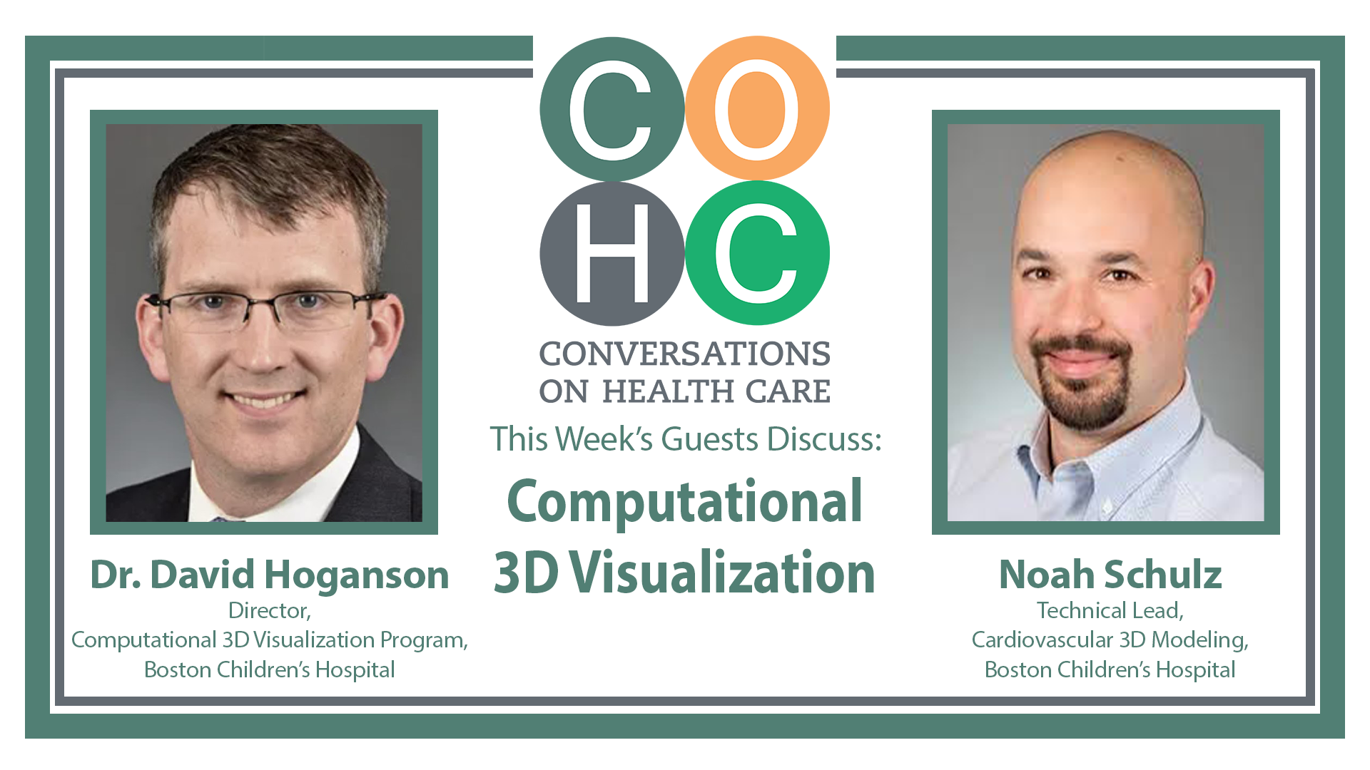
One of the first steps in creating your “digital twin” is already taking place at innovative medical centers across the U.S. Boston Children’s Hospital and 20 other centers like it are using 3D cardiac models to improve pediatric heart surgery and help families better understand their child’s care.
The 3D visualization program creates patient-specific digital replicas of pediatric heart anatomy that surgeons use to plan complex procedures. The models have also become a powerful communication tool, helping parents see what is happening inside their child’s heart.
“We want the families to be part of the decision-making about what do we do and what [are] the next steps for their children,” says Dr. David Hoganson, director of the Computational 3D Visualization Program at Boston Children’s. “Without really a clear picture, often they’re just left to ‘Whatever you say, doc,’ and that’s not really the best option.”
Last year, the Boston Children’s program made 3D models for roughly 60% of patients going to the operating room.
“We’re pooling our collective efforts … to help really be the rising tide that lifts all boats and really sort of do this together as a community,” Hoganson tells hosts Mark Masselli and Margaret Flinter.
Noah Schulz, the program’s technical lead, highlights future opportunities. “If we can get to the point where … my colleagues can spend more time doing the complex surgical planning work and communications work that we know is really hard to replace with AI, I’d be happy with that,” Schulz says.
For Hoganson, the journey is personal. As a child, he had ventricular septal defects.
“It’s really gratifying as a person to take care of these children who are in the position I once was,” he says. “And it’s really been fun to help build solutions that address what I had and try to make that a more reliable outcome for these children.”
(00:00)
[instrumental music plays]
Mark Masselli (00:04)
Are you ready to meet your digital doppelgänger? More than just a novelty, this kind of 3D visualization is a
innovation, improving diagnosis, surgical planning and outcomes.
Dr. David Hoganson (00:16)
There's a whole spectrum of what, digital twin encompasses. But, you know, I think for, for our clinical needs,
really the focus is, you know, what's the child's anatomy and physiology going into the operating room, and how
do we correct that, you know, with the surgery we do. And, and, you know, and doing that in a very
computationally intense and pre-planned way is, is a reality right now. So yesterday at Boston Children's
Hospital, we did five cardiac operations, and four of them, had computationally pre-planned, either patches or
valve repair. And, and that was an integral part of, of that for four of those five children.
Margaret Flinter (00:58)
And our guests are Dr. David Hoganson, the director of the Computational 3D visualization Program at Boston
Children's Hospital. And Noah Schultz, an engineer who specializes in segmentation and virtual surgery.
Dr. Hoganson (01:12)
I'm a patient as well as a surgeon, so I had complex congenital heart disease. I had multiple muscular VSDs,
and, and, underwent two surgeries as a small trial to repair that. This was before the days of CT scans and 3D
models. This was actually before the days of echo, which is hard to, believe. and, you know, I'm, I'm incredibly
grateful for the care that I received and the opportunity to normal life. And, you know, in fact, if, if I was a
patient in Boston Children's today, I would have a 3D model and, and virtual surgery planning. And it's really,
you know, it's gratifying as a person to take care of these children who, you know, are in the position I once
was. And it's really, been fun to help, you know, build solutions that, that, you know, address, what I had and try
to make that, a more reliable outcome for, for these children.
Margaret (02:00)
This is Conversations on Healthcare.
Mark (02:13)
Well, Dr. Hoganson and Noah, welcome to Conversations on Healthcare.
Dr. Hoganson (02:17)
Thanks so much for having us, really appreciate.
Mark (02:19)
Yeah. Well, let me just say at the beginning, we, we had the opportunity to see you at Aspen Ideas Health, was a
really incredible presentation, but we're in primary care. But we were so enamored with your presentation. We
thought we'd, have you on and have this conversation, you know, your, doctor, internationally recognized expert
in digital modeling, modeling and computational 3D visualization program for surgeons. I'm, I'm wondering if
you could just tell, walk our audience through, what that is and why it's so important, to the industry.
Dr. Hoganson (02:59)
Yeah. I mean, you know, for children with complex congenital heart disease, the challenge, you know, that we
face in repairing them surgically is to, is to really reconstruct the anatomy, to, to achieve really, normal blood
flow through the heart and the blood vessels. And this often evolves either patch enlargement or downsize of a
blood vessel or redirection of blood flow within the heart. And, you know, for the last several decades over
which these techniques have evolved, it's really, you know, the responsibility of getting those patches just right
has, has been based upon the intraoperative, visual estimation of, by the surgeon. And, you know, these are
really complex three dimensional structures, and getting that right in real time, has, you know, has been a
challenge. And so really, we, we started this whole program, with an effort to, to really take an engineering
approach to doing that and pre pre-planning and predetermining what those patches and those reconstructive,
surgeries should entail. And then carrying those plans into the operating room. And the goal is to improve the,
both the geometric outcome, but really the clinical outcome for these children. So, they have as good of a
physiology as they can and, and grow and live, you know, normal and, and healthy lives as much as possible.
Margaret (04:23)
Well, any, parent who's facing cardiac surgery for their child is of course, in terror, right? As they, right. Think
about what needs to be done. And you introduce something like this, which I'm sure you have the data, to,
suggest how much, more effective or safer it is, how do you, how do you deal with what we call informed
consent? How do you educate and inform parents about something which is almost impossible to imagine? Have
you developed a library of video content, or is it, storytelling? Tell us a little bit about that process.
Dr. Hoganson (04:52)
Yeah, you know, I, I, I think, I think from the parent perspective, you know, certainly, you know, as a parent of
a child facing, you know, a life-threatening condition, the needs of major surgery, you, you know, you, as Noah
has said in the past, you want no stone unturned, right? You want everything possibly done to prepare for that
operation and conduct that operation in a way that you'll have a, a predictable and outstanding outcome, right?
And so, I think our, our interactions with the parents, you know, when they, when they see the work we're doing
and, and this, you know, sometimes substantial additional effort of a whole team of engineers is really one of
gratitude. You know, just thankful that all this, you know, work and planning is being done to really make this
as successful as possible. So that, that has really been the very consistent feedback. not only are these models
useful for the surgeons and the cardiologists, but they're really useful as a communication tool with the families.
You know, this is really, you know, often we're facing very complex heart disease, and we want the families to
be part of the decision making about what do we do and, you know, and, and what's the next steps for their
children. And, and without really a, a clear picture, often they're just left to whatever you say, doc. And that's
not really the, the best, the best option. So we, you know, when we make these 3D models, we share them with
the families, and, and it has transformed that experience with many of these families, to be able to, you know,
sometimes for the first time well into the steps of care for their child, understand what the features of their
child's heart disease are, and really understand what the steps are gonna be taken to, to repair that. So, it's really
been a phenomenal educational tool and where we continue to expand, the availability of these models to help
parents understand, you know, the, the details and the, the plans to repair the heart disease.
Margaret (07:13)
Well, I think that's fantastic because as we know, even the most highly educated people often are completely
clueless about the anatomy of organs, like the heart, so to do this is so important.
Mark (07:04)
And even they say, “Okay, doc, whatever you say.”
Margaret (07:08)
And even- right. Exactly.
Mark (07:09)
So, is this robotic assisted or, or, or remote telesurgery? What- how, how's the surgery done?
Dr. Hoganson (07:18)
Yeah. So, the, the, for pediatrics, it's all essentially done by hand. Okay. there's not really a robotic or
telesurgery role, but I think what, what is different is in the operating room, we use these 3D models extensively
throughout the operation. So, if you come in my operating room, you'll see the 3D model up on the screen on
several screens in the operating room, and then the details of the patches that we planned and all that is available
in real time. We have a sterile controller that we can control the model from the field and, you know, and use it
throughout the operation. And, and in many of these complex surgeries, it's really essential, to have that
information and all the spatial relationships of, you know, often the, you know, the congenitally different
structures and the different place of those have all that information in real time has really been an, an
outstanding step forward.
Margaret (08:09)
Well, one of the points that I think you, might have made when we heard you speak, was the reality that
sometimes reintervention or redoing this, the cardiac procedures is a real factor, and this is less likely to happen.
Tell us more about that.
Dr. Hoganson (08:25)
Yeah, yeah. And that's, and that's really one of the targets of this effort. You know, this is not about making it
easier on the surgeon. It's not really about reducing the or time. This is about avoiding, complications and, and
reinterventions. And in some of what we do that, that can be as high as 25 or 40%, reintervention rate with the
current way things are done. This is just, it's complicated geometry, it's complicated problems, things have to
grow. And, and, you know, we know if it's, if it's done really well the first time that that, that rate of
reintervention is much less. And so, we, we've seen that in the things we've done, and some of the things that
we're tackling right now and about to start using clinically are some of those really high reintervention rate
situations. trying to reduce that with really precise, preoperative planning.
Mark (09:14)
Yeah. Noah, how challenging is it to create this 3D model? And, and I'm wondering if modeling is being used
for other surgeries beyond heart surgeries, or could it be, and I guess we wouldn't have an interview without
asking if AI is involved, in this process at all.
Noah Schulz (09:34)
Yeah, that, so there's a, you know, there's a long recent history of the 3D modeling, anatomical 3D modeling
being used for surgical planning, not just in cardiac surgery, but more broadly in orthopedics and general
surgery. And, quite often in plastics and, you know, craniofacial surgery. And, I've been at, at, Boston Children's
Hospital since 2014, and the first few years I was making printed 3D models for a number of surgical specialties
for, for surgical planning, for communication with patients and families and, and other things. And so, it, it, it's
broadly applied, but I think, what we found, at least in cardiac surgery is, that the, you know, the variations in
anatomy are great, and the, the differences right between the open heart during the procedure and the heart at
normal physiologic pressure are, are pretty great as well. And so having access to that 3D anatomy and those
relationships is just hugely valuable as, as Dr. Hoganson said. And I think, you know, the complexity of, model
making has, has lowered as the imaging quality has gone up, even over the last, you know, dozen years that I've
been doing this, I've seen improvements in, in, you know, three-dimensional imaging, that we use as the basis
for these models. And, that makes, that makes our job a little bit easier. Of course, we're applying these more
broadly to more challenging anatomies and more challenging diagnoses. And so, the anatomical sophistication
goes up. And so, the, the pressure for myself and for the other engineers that I work with to learn cardiac
anatomy, even, you know, even more, than we already have goes up. but the tech, you know, that we've seen a
lot of technical improvements. artificial intelligence is, you know, and, and sort of image processing is being
used in some surgical specialties, particularly, like I said, in orthopedics. I think it's a little bit easier to apply.
we've seen some of these tools, and there are plenty of, you know, both sort of commercial organizations and
researchers working on developing AI segmentation tools for cardiac anatomy. But the challenges are pretty
great. And so, what we've seen is it's the early stages I expect. And, and honestly, if, if we can get to the point
where these tools are really effective, and I can, I am, and my colleagues can spend more time, doing the
complex surgical planning work and communications work that we, that we know is, is really hard to replace
with AI, I, you know, I'd be happy with that. I'm not there. It's still very hard.
Mark (12:15)
You know, I’m gonna draw on my, I'm gonna draw on my Star Trek knowledge. Is this something that could be
a hologram, that you could walk around that, that a, a physician could walk through this model? Or is this
something, you know, how, how does Dr. Hoganson look at this? Is it just on a screen or how is it visualized?
Noah (12:37)
Yeah, we're experimenting. We're always looking at new visualization technologies. There's, you know, some of
these models can be viewed in VR, you know, using a regular VR headset that you might use for video games.
there are holographic 3D displays, both that have special glasses like you might wear at the movie theater or
using other technologies that allow you to have some of that spatial depth information encoded. But often, at
least in the operating room, it's a standard, you know, 4K color display. So, you, you get some sense of the
depth information and obviously manipulating the model gives you a lot more of it. But, we're always on the
lookout for better ways to experience these things. And, we, you know, we've got a number of things on the
horizon that that'll hopefully make it more accessible.
Margaret (13:24)
Well, it sounds very complex and also like you're at the epicenter of innovation, but I understand that you also
use some tools that our audience might be familiar with, such as a, a Wii game controller. So where does that fit
into the mix? I can't quite imagine somebody guiding this with a game controller.
Noah (13:43)
Yeah, that's, that's exactly right. So, you know, early on in this process, as we moved into the operating room
with these models, surgeons like to be able to drive on their own. They're, they're, very independent minded. and
so, there was a, there was a, you know, a few years where I would just sit in the corner of the operating room
with my ear, you know, stretched out, waiting to hear a surgeon say,” Hey, can you rotate that a little bit to the
left?” Or, “Can you zoom in a little bit on this thing?” And, you know, obviously that's like a pretty inefficient
use of resources. So, we, I went, I went home and I borrowed a Nintendo controller from my kids and, brought it
to work, put it in a sterile bag. And we have been using that, you know, the surgeons have been able to use that
from the sterile field using, using a couple of tricks to allow them to drive, basically, so they can more easily
manipulate these things and, and preset some views. So, as they work through the case, they can have access to
the information that they've already, you know, provided to the model or, or measurements that they've already
made.
Mark (14:47)
That's great. Dr. Hoganson, as I mentioned at the top of the show, we heard you at Aspen Ideas Health, about
the ultimate in personalized medicine, the digital twins. I'm not sure if that's a Apollo reference, or not. I think it
has some historical reference, that could predict chemotherapy responses, I think determine pregnancy outcomes
and really help design custom tailored prosthetics. I'm wondering how far along are these concepts from
becoming reality.
Dr. Hoganson (15:18)
Yeah, I mean, I, I think there, you know, there's a, a whole spectrum of what on Digital Twin encompasses, but,
you know, I think for, for our clinical needs, really the focus is, you know, what's the child's anatomy and
physiology going into the operating room, and how do we correct that, you know, with the surgery we do. And,
and, you know, and doing that in a very computationally intense and pre-planned way is, is a reality right now.
So yesterday at Boston Children's Hospital, we did five cardiac operations. Hmm. And four of them, had
computationally pre-planned, either patches or a valve repair. And, and that was an integral part of, of that for
four of those five children. So, it is a reality. Now, last year, we made, 3D models for almost 60% of our, of our
patients going to the operating room and, and virtual surgery planning including, flow simulation with the same
software they use to design airplanes to say, what's the flow gonna be through the heart after the repair? And,
and, you know, we often use that to select which operative strategy to use. So, so it it's a reality now here and,
you know, we're working, with, over 20 centers around the country to help sort of them expand this. and so, it's,
you know, it's available to, to, you know, someday all patients who, who could benefit from it going, going
through surgery.
Margaret (16:45)
Well, Dr. Hoganson, I know, from knowing our audience that people would probably like to hear from you a
little bit about the evidence base that you've developed in terms of outcomes. You talked about, lower need for
reintervention using this approach. would you like to comment anything from infection, outcomes, length of
stay, whatever, whatever you look at that you have found important?
Dr. Hoganson (17:08)
Yeah, so, you know, I think, I think it's, it's both a combination of, you know, where, where we are without this,
you know, sort of what's the current, you know, approach and what's the current results, and then, and then what
can we achieve with this? And so, you know, we've seen anywhere from tackling, you know, surgeries that have
either several standards of deviation of sort of size variation of what we do from a patch planning or have a very
higher intermission rate. And we've dropped that down dramatically. and for other patients, particularly those
that we've done the flow simulation on, these are patients often who have to come back to surgery because the
way the first surgery was done did not result in adequate physiology. And we've been able to correct that or in
other patients, completely avoid that. And in, in some patients, for example, they have about a almost a 75%
chance of develop of needing a reintervention. And in our series, we've reduced that to zero, with this, flow, the
flow simulation testing. So, it's really quite remarkable. And in, and in children, it's both, it's both a risky
endeavor to go back to the operating room. And it's also very expensive, of course, you know, these are, these
are operations that have ICU stays after and then, you know, stays weeks in the hospital. And, you know, in one
evaluation we did for the flow simulation, we, we've, saved hundreds of thousands of dollars per case that we've
evaluated just because we reduced the risk of a, a very expensive, you know, a fraction of a million dollars sort
of reintervention. So, it's, it's really, very impactful. But I think the, the, the most important thing is for the
patients, right? For these children to not have to go back to the catheterization lab, not have to go back to
surgery and, and be able to have as normal of heart function and exercise, capacity as possible. And that's the
real, that's the real target. And, you know, we're continuing to, to build that data, particularly as these patients
get older and we can start to run exercise tests and those things and really show, the, the impact of this.
Mark (19:14)
Well, it begs the question of, of the use of these 3D modeling for complex cases has grown from, as you were
mentioning, just from a small fraction to over a quarter of major surgical cases over a few years. How do we get
to increase it even more?
Dr. Hoganson (19:29)
Yeah, so I mean, I think, you know, I think part of, part of this has been the, the technology piece and the
integration with the clinical care. and part of it is, is how do you make this part of the standard clinical practice,
right? And, and one of the pieces of that that's really important is, you know, is that this is reimbursed in a, in a
reliable way. And so, our team has tackled that also. And we just, this fall got a reimbursement code approved
as CA new CPT code approved for these efforts. And that's been a huge piece of this. So, and just integrate this
into the normal part of billing and reimbursement. And so, engineers like Noah, you know, are, you know, their
efforts are reimbursed as part of the standard medical care. And that's, that's been a big, a big push for us. And,
you know, I think it's really enabled other centers to, to take this on, knowing that there's, you know, there's a
way to integrate this into the standard, you know, reimbursement and, and medical care.
Margaret (20:25)
Well, one of the things that, brings to my mind is that you can't do this without a incredibly sophisticated and
well-trained workforce. We're always, concerned with the democratization, if you will, of healthcare. You
know, how do we have it be available to everybody. Maybe you could share with us a little bit, most, surgical
teams in, back in the olden days of, oh, six months ago, didn't have, didn't have engineers, at the controller off,
to the side or other, innovations that would just come at a pretty dizzying speed. What are, what is your
institution doing around workforce training, fellowships, the technicians, the nurses that, really support this kind
of intervention? The surgeon is key, obviously, but takes a whole lot of people on the team to make this work.
Dr. Hoganson (21:13)
Yeah, it, it, it really does. You're, you're, you're right. And it's been a, a journey for us to, to grow our team. you
know, I think, I think the, the part of what we've, integrated into all of this is, is how do we simplify this so it
can be done with a, a much smaller team than we have, you know, and, and, you know, one or two engineers per
center that can tackle, you know, most of this. And so, we've kept that in mind with all of these different
workflows we've developed and new clinical approaches and, and now we're in the process of training other
centers who've hired engineers and are really, modeling our approach. And it's, it's been exciting to see that
grow and, and work just as well at other centers as it has, for us. And so, I think it's, it's, a simplified model that,
you know, many other centers are taking on, and it's, it's exciting to see it start to impact patients, all across the
country.
Mark (22:02)
Dr. Hoganson, I, I think our audience would love to hear from you, if you wouldn't mind sharing your personal
story, and your history with complex congenital heart disease and it, and its effect on your life.
Dr. Hoganson (22:16)
Yeah. I'm a patient as well as a surgeon. So, I had complex congenital heart disease. I had multiple muscular
VSDs, and, and, underwent two surgeries as a small child to repair that. This is before the days of CT scans and
3D models. This was actually before the days of echo, which is hard to, believe. and, you know, I'm, I'm
incredibly grateful for the care that I received and the opportunity to normal life. And, you know, in fact, if, if I
was a patient in Boston Children's today, I would have a 3D model and, and virtual surgery planning. And it's
really, you know, it's gratifying as a person to take care of these children who, you know, are in the position I
once was. And it's really, been fun to help, you know, build solutions that, that, you know, address, what I had
and try to make that, a more reliable outcome for, for these children. So, I'm, I'm in incredibly grateful to be in
the position I am to make a difference. And, it's been a, a very rewarding, piece of all of this.
Mark (23:16)
You know, you were talking earlier about, 20 other centers maybe, share sharing information. What's that
network look like? does it have a umbrella organization or, I think people would be, interested in knowing more
about, the collective work that's going on in this area?
Dr. Hoganson (23:34)
Yeah, it's, it's, it's really evolving quickly. And so, we're, in fact, we're creating that umbrella organization right
now, that sort of is a collaborative network among these centers. and, you know, several great institutions across
the country have been tackling this also, and now really pooling our collective efforts in places like Children's
Hospital, Philadelphia and Cincinnati Children's in Stanford and Columbia, and, and really putting those efforts
together to help, you know, really be the rising tide that lifts all boats and, and really sort of do this together as a
community. and it's, and it's been fun to take that approach. And, we're excited to see, how this will continue to
evolve and expand across the country.
Mark (24:39)
It's a difficult business. The children's hospital world. I, I know there's been some consolidation, large
benefactor has been, you know, trying to organize, children's hospitals all across the country.
Margaret (24:27)
Well, and children's hospitals, of course, the whole GME funding formula has always been problematic. But I
know there's been a lot of progress on that. And, and I think, not just across the country, but certainly what
you're doing is a global opportunity for us to make things better. And, you know, Dr. Hoganson, I appreciate
your sharing your personal story, and I'm reminded one of our wise, physicians here in our organization, always
saying the best, the best doctor for you is the one that has the same health problems you do. They're really
fascinated with how we, how we approach those.
Dr. Hoganson (25:00)
Yeah, no, for sure. It's, it's a, it's a passion project for me of, of course. And, you know, we, we have been, you
know, able to be involved in several, you know, centers around the world. And in fact, we're making a model
right now. I think Noah's making one today for a center in Africa. Wonderful. And we've, we've worked all
across Europe and, and South America, and just tried to, you know, again, help, help these centers, you know,
tackle these hard problems in a similar way that we have. And it's been fun to be, to be part of that both here,
here, and abroad.
Margaret (25:29)
Perfect. Well, my, my, next question was going to be the, the next leap. Is there a place for fully autonomous
robots performing cardiac surgery in the near future? Or is that, still way off or maybe never to be?
Dr. Hoganson (25:44)
Yeah, I mean, I, I, I think what's next for us in the, in the, the challenge we're tackling is how do you really,
make these workflows happen much quicker and, in some ways, in a semi-automated way. So, the, the patch
planning and the pre-planning that we do that currently can take tens or, you know, 50 plus hours per patient,
how do we make that so quick and still as reliable that it can be applied in many, many, for many, many other
patients? And, you know, I think that's what we're seeing in the next several years happen is how do we take the
guesswork, which is currently still the gold standard, and sort of that intraoperative visual estimation, how do
we make that a, a pre-planned standard, and, and reduce the variability and improve outcomes, you know, you
know, across the board. So, those are the things we're tackling and, we're excited to see, you know, what comes,
Mark (26:44)
You know, Dr. Hogan said, I think our audience would be interested to know that at one point you were testing
a procedure using veins from a baby's own umbilical cord to stabilize blood flow in the hearts of those born with
complex defects. Tell us what's the latest on that effort?
Dr. Hoganson (26:54)
Yeah, you know, I think it was, it was an important solution to the, the problem that we faced at the time where
a lot of these patients were getting these small plastic tubes, and they would, and they would clot off at a, at a
really, remarkable rate. thankfully, you know, as things have evolved, the same stents that we use for adult
coronary artery disease are now able to be used in babies. And instead of putting in the plastic tubes by and
large, we put a stent in the ductus arteriosus, to accomplish the same thing. So, so the rate of using those small
plastic tubes has gone to not zero, but close to zero. And so, that effort has really been replaced by ductal
stenting, which is, which is wonderful for the patient because the recovery is quicker. And, and that's, that's been
an important step forward for the, for the field. So, you know, I think, you know, all everything we do will
continue to evolve and we're, looking forward to watching the same thing happen with, you know, this 3D
modeling effort.
Mark (27:49)
And, and just on the federal support, how's the NIH been in your efforts? Are they supportive of the work that
you're doing?
Dr. Hoganson (27:58)
Yeah, they really are. And they've been supportive of our group and, you know, and other, congenital cardiac
groups across the country. And as you mentioned, there are other, other nonprofit, you know, benefactors
who've stepped in for patients with single ventricle heart disease or, or broadly across congenital heart disease.
And I think all of those resources are really important for our field, to really, you know, take these steps
forward. These are, these are small patient volume, you know, high risk scenarios, and, you know, having all
these, different, avenues of support is really important as we try to, you know, make advances for these children.
Margaret (28:36)
Well, we want to thank you Dr. David Hoganson, Noah Schultz, for the incredible work that you're doing and
for joining us today. And thank you to our audience for being here. Just a reminder to be sure and subscribe to
our videos on YouTube. Find us on Facebook and X with our account name, CHCRadio. And as always, please
go online to chcradio.com and sign up for email updates. And please share your thoughts and your comments
about this program. Take care and be well.
(29:01)
[instrumental music plays]
Margaret (29:04)
This copyrighted program is produced by conversations on healthcare and cannot be reproduced or remitted in
whole or in part without the express written consent from Community Health Center, Inc. The views expressed
by guests are their own, and they do not necessarily reflect the opinion of conversations on healthcare or its
affiliated entities.
(29:26)
[instrumental music plays out]